A DX-Based Dose-Reduction Program
Digital Transformation for Radiation Protection
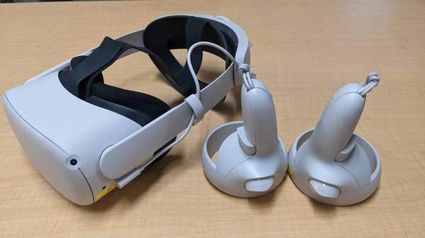
Because radiation is invisible, it is difficult to intuitively grasp the effect of protective actions. Using digital technologies such as AR/VR/MR (XR), our laboratory visualizes radiation scatter and dose distribution so that radiological staff can experientially learn “where to stand to reduce exposure.” As clinical research on occupational diseases, we also verify the effectiveness of these materials.
XR Visualization Materials
Spatially visualize scattered radiation and dose distribution with AR/VR/MR to learn protection principles intuitively.
Action Checklists
Organize dose-reduction actions by examination and department to support practice in the field.
Training Radiological Technologists
Radiation protection education in the School of Health Sciences and graduate programs, fostering the next generation.
Courses Taught
Main teaching areas
| Undergraduate | School of Health Sciences, Department of Radiological Sciences (Radiation Management, Radiation Therapy Technology, etc.) |
|---|---|
| Graduate | Graduate School of Health Sciences, Field of Medical Quantum Science (Radiation Protection, etc.) |
| Center | Center for Integrated Radiation Safety Management (concurrent; safety management education) |
Occupational Exposure of Healthcare Workers
Why radiation protection education is needed
As radiological practice increases, not only radiologists but also physicians and nurses in cardiology, gastroenterology, neurosurgery, orthopedics and urology are exposed during X-ray fluoroscopy procedures. The risk of cataracts from lens-of-the-eye exposure has drawn attention, and in 2021 the equivalent dose limit for the lens was lowered.
We surveyed the lens exposure of 4,493 person-years of radiological staff in National Hospital Organization facilities and showed that physicians and nurses engaged in fluoroscopy are relatively likely to exceed the dose limit (20 mSv/year) (Fujibuchi et al., 2021).
Three Principles of Protection & Protective Equipment
Time
Keep X-ray exposure time to a minimum; dose increases in proportion to time.
Distance
Keep as far as possible from the source; do not put hands in the field (dose differs >10× inside vs. outside the field).
Shielding
Place shields/protective gear between the scatter source and the worker; optimize equipment QC, exposure conditions and collimation.
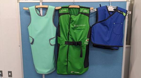
Protective equipment includes wearable types (aprons 0.25–0.35 mmPb, eyewear, thyroid shields, gloves) and non-wearable types (shielding plates, curtains, screens). We verify their effective placement with scatter simulation.
Three Pillars of the DX-based Dose-Reduction Program
🌐
1. Education Portal Site
A site bundling text, images, videos, webVR materials and action checklists for interactive learning, with efficient analysis of comprehension.
🧐
2. 3D Radiation Visualization Materials
Simulation-based VR/AR visualizing scatter distribution; AR apps for cardiac cath, ERCP, CT and mobile C-arm.
⚠
3. Real-time Exposure Warning
Linked with wireless dosimeters and scatter-visualization cameras to estimate and warn of operator exposure in real time.
Scatter Visualization AR App “X-SERVE”
Our scatter-visualization AR app “X-SERVE” (X-ray examination Scattered Radiation Visualize application) comes in three variants:
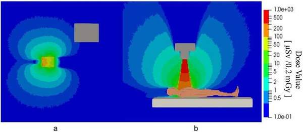
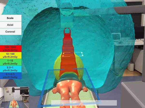
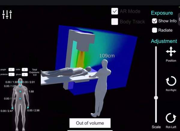
• X-SERVE: visualizes scatter in CT and similar exams in 2D/3D with distance and dose from the field center.
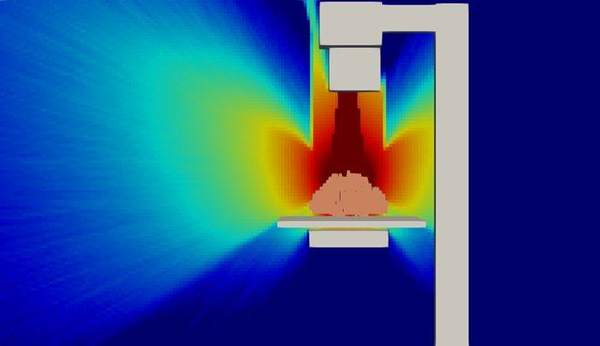
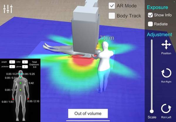
• X-SERVE IVR: for C-arm fluoroscopy; observe how the scatter distribution changes with angle and shield placement.
• X-SERVE VD (Volume Data): shows dose distribution on arbitrary cross-sections, staff dose and dose at the camera position, and compares cases with and without a protective curtain. Supports AR / 3D-viewer modes and body tracking.
Related Project Site
The outcomes of our radiation protection education are also published on the project site below, offering materials on the importance of radiation protection, the reality of occupational exposure, dose evaluation and monitoring, department-specific protection measures, and action checklists.
Videos
Demonstrations of scatter visualization and dose evaluation (from our YouTube channel “lab fujibuchi”)