Visualizing "invisible" radiation in clinical settings for safer radiological practice
While radiation is indispensable for medical diagnosis and treatment, it also entails the challenge of exposure to patients and healthcare workers. Built on radiation protection science, our laboratory aims to realize safe, high-quality radiological practice through dose evaluation and monitoring, radioactive waste management, and the visualization of radiation combined with XR-based educational materials.
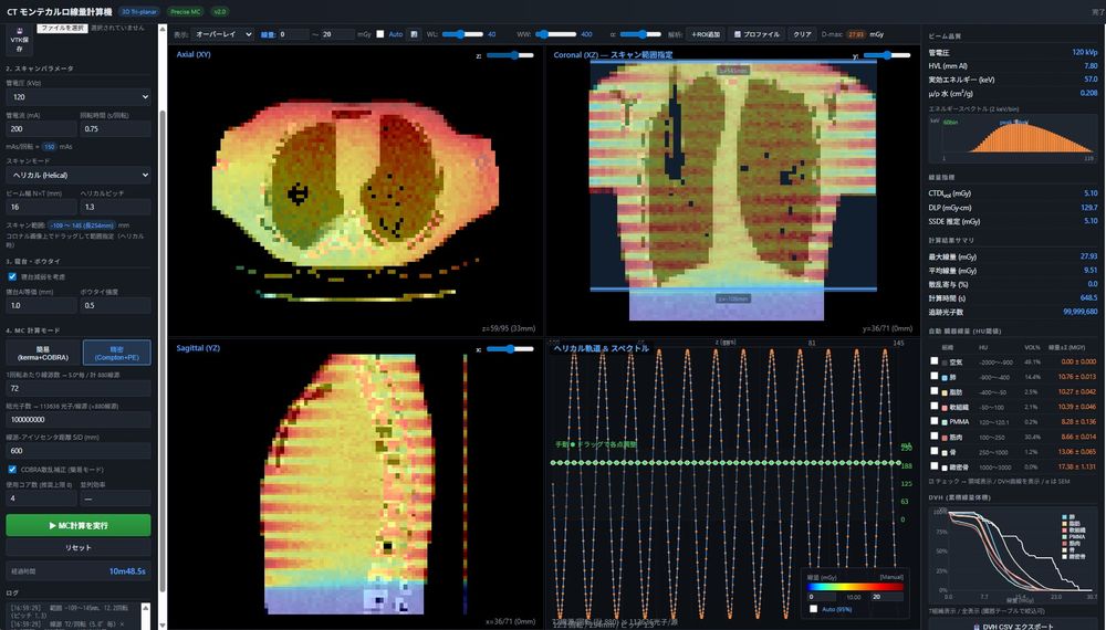
We assess exposure by measurement with dosimeters and phantoms and by Monte Carlo simulation, and work on optimizing medical exposure with image quality in mind, dose management, monitoring of healthcare workers, and dose-reduction measures using digital technologies, including performance evaluation of protective equipment.
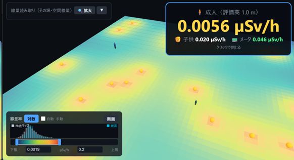
We evaluate radioactivity and dose in contamination surveys, in activated components of medical accelerators and in radioactive waste from nuclear medicine, as well as the effect of decontamination after nuclear disasters.
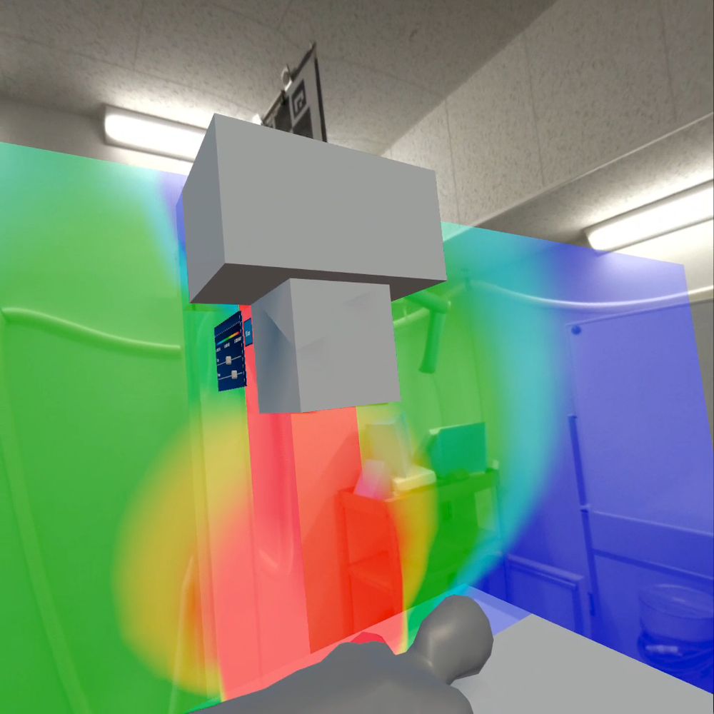
We visualize "invisible" radiation with AR/VR/MR (XR) and implement it as radiation-protection teaching materials. We develop DX-based dose-reduction programmes, build them into lectures and practical training, and go on to verify their educational effect.
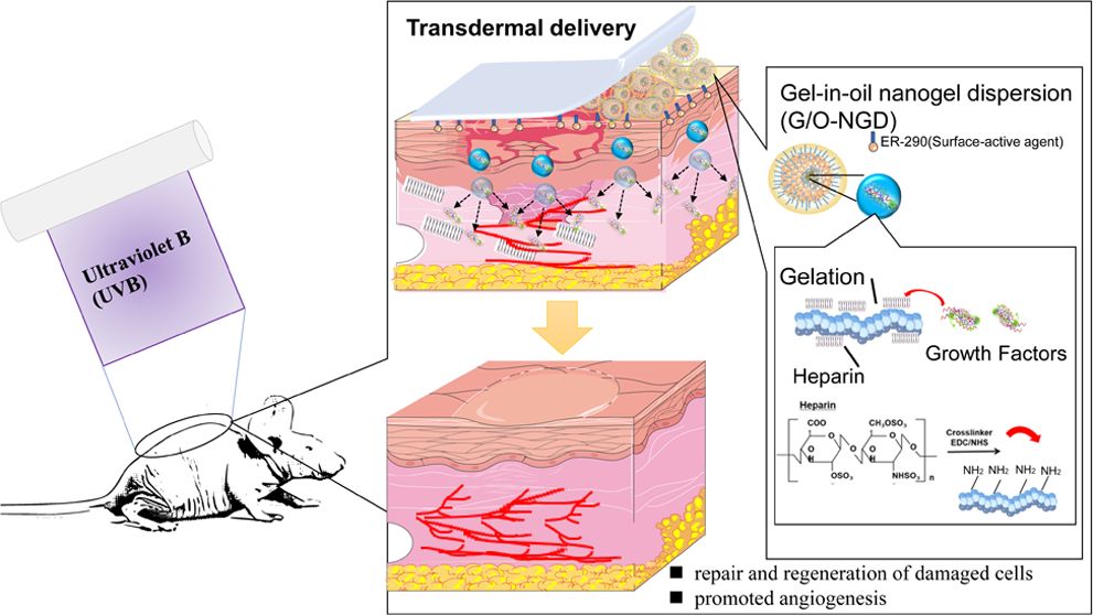
Through cell and mouse experiments, we develop protective agents against radiation dermatitis and evaluate their effectiveness.
Figure: a mouse study on preventing and repairing ultraviolet-B (UVB)-induced skin damage (Zhang et al., ACS Omega, 2023; Fujibuchi is a co-author).
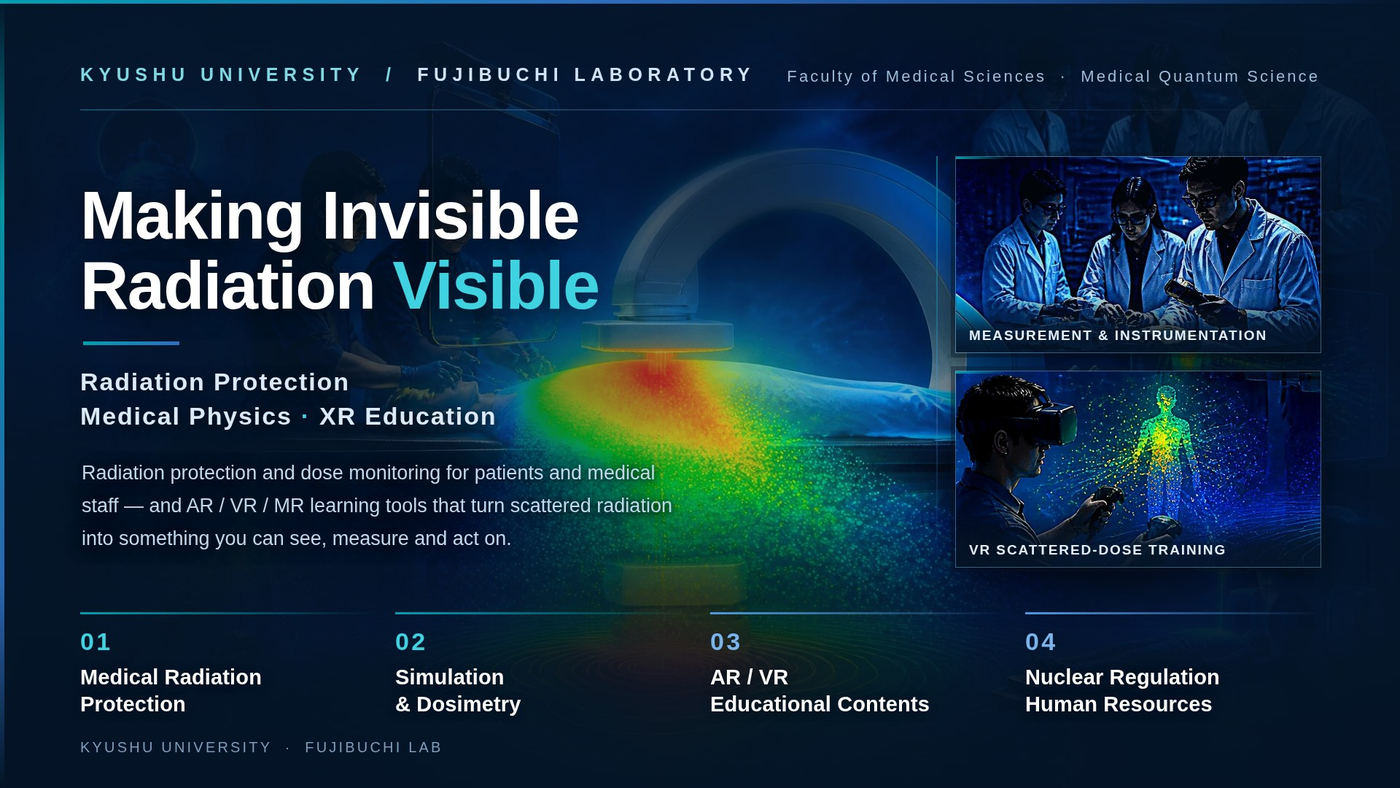
Externally funded programmes led by our laboratory

Selected jointly with Tohoku University under the Nuclear Regulation Human Resource Development Programme in FY2021 (to FY2025). Through laboratory practice in radiation management, facility visits and internships, the programme trained people who can put radiation protection into practice. It ended in FY2025 and continues as a follow-up project from FY2026.
FY2025 final report (Nuclear Regulation Authority, in Japanese)

From FY2022 to FY2024 we carried out “Development and evaluation of the effectiveness of a dose-reduction programme for medical staff using digital transformation”, funded by the Industrial Disease Clinical Research Grant, and developed and published practical materials such as visualisation and sonification of radiation for hazard prediction, department-specific protection measures and action checklists.
Research reports (Ministry of Health, Labour and Welfare; PDF, in Japanese)
Show the invisible, so that protective behaviour becomes second nature
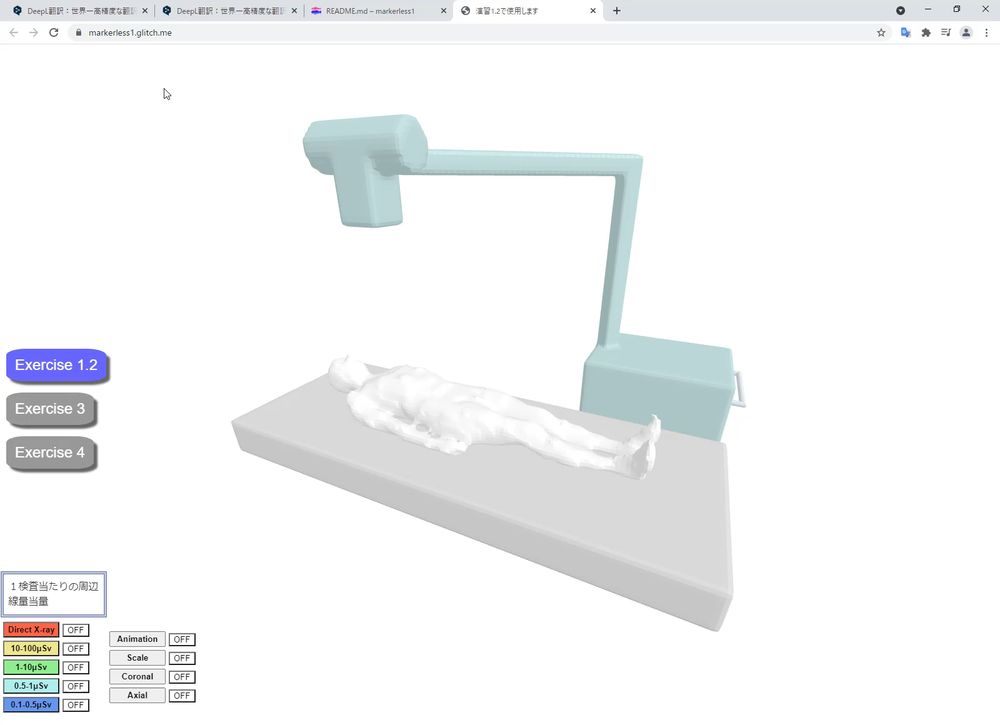
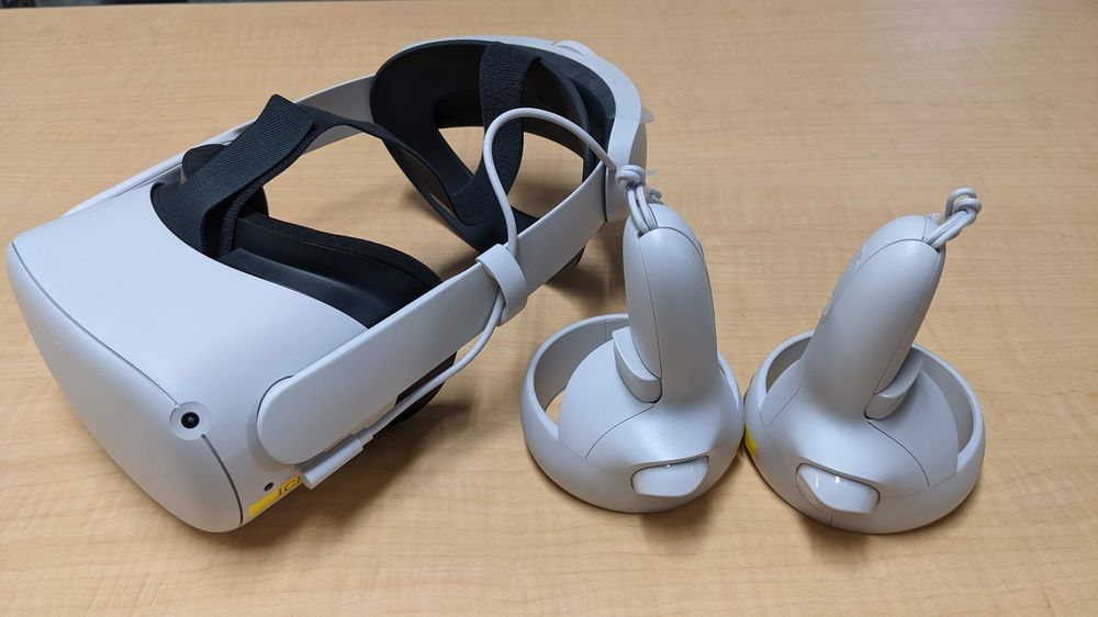
No VR headset required. Observe the scatter distribution from any direction on a PC or smartphone. Also used for remote practical training.
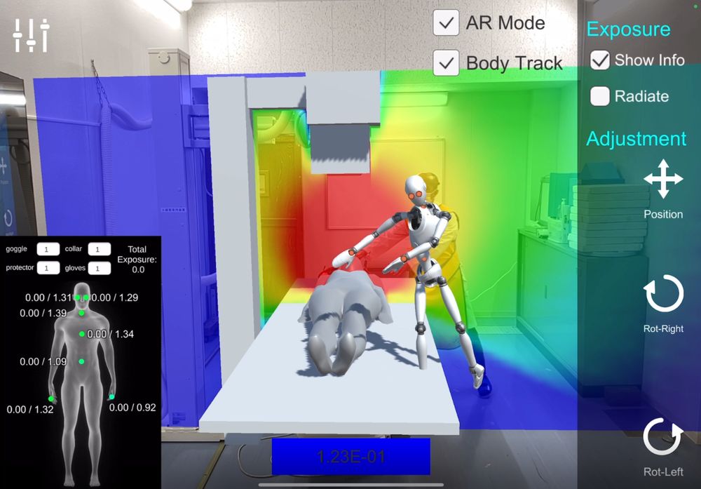
A portable AR application for iPad that overlays the 3D scatter distribution onto the actual examination room, so learners feel how position changes exposure.
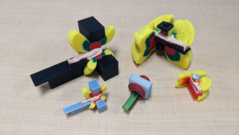
Scatter distributions printed as physical objects. No device or power supply required, at a material cost of roughly two US dollars each.
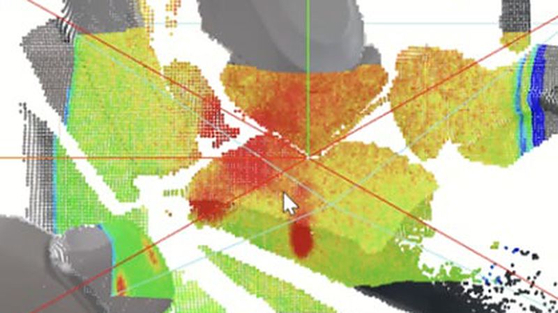
We measure scattered radiation with a camera that combines a pinhole and a scintillator, and with the compact Timepix3 semiconductor detector, and turn where it comes from and how it spreads into images.
Figure: Kamochi et al., Applied Sciences, 2026 (DOI, CC BY 4.0, cropped)
Our lab introduction and demonstrations from our YouTube channel
Research activities and results, updated regularly
Most of our news is posted in Japanese; the latest posts are listed below (in Japanese).
(in Japanese)
(in Japanese)
(in Japanese)
(in Japanese)
(in Japanese)
(in Japanese)