Integrating radiation protection, monitoring, and visualization technologies
This page presents our laboratory's research into making radiation protection work in real clinical settings. The underlying ideas (the ICRP principles, and medical, occupational and public exposure) are explained in Basics, and our teaching materials in Education.
Our research rests on two pillars: "radiation protection, monitoring, and radioactive waste of patients and staff in medical radiation use" and "visualization of radiation." Combining experimental measurement, numerical simulation, and field surveys, we propose evidence-based strategies for dose reduction.
Four methods combined into protection strategies that work in the clinic
We measure exposure with dosimeters and anthropomorphic phantoms on real equipment under clinical conditions, and survey actual practice in hospitals.
Monte Carlo simulation gives us organ doses and room-scale scatter distributions that cannot be measured directly. We validate the results against measurements.
We render the resulting dose distributions in XR (AR/VR/MR) and as CG models, so staff can grasp protective behaviour intuitively.
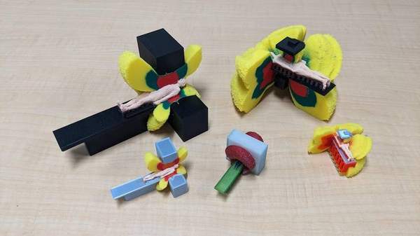
With 3D printers we build phantoms whose CT numbers match human tissue, and physical models of scatter distributions for teaching. We design and make both the tools we measure with and the tools people learn with.
These four are not separate activities but a cycle: measurement validates simulation, simulation provides the evidence behind our visualization materials, and the questions raised by visualization drive the next round of measurements. The phantoms and models we 3D-print support both measurement and teaching.
Medical use of radiation continues to expand, and Japan — with a very large number of CT scanners per head of population — is considered to have a high level of medical exposure by international standards. At the same time, the 2011 ICRP Seoul Statement (formally published as ICRP Publication 118, 2012) substantially lowered the equivalent dose limit for the lens of the eye, making occupational dose management for staff in interventional radiology (IVR) an urgent issue.
Yet radiation is invisible and gives no sensation, so protective behaviour is hard to sustain. Solving this "invisibility" with technology is the consistent theme of our laboratory.
To optimize medical exposure by weighing patient dose against image quality, we study dose evaluation and dose management systems. In addition to measurements with dosimeters and anthropomorphic phantoms, we obtain organ doses and dose distributions that cannot be measured directly by Monte Carlo simulation, and validate the calculations against measurements (see Precise Dose Evaluation for Medical Exposure below).
We assess the exposure of healthcare workers in fluoroscopy/IVR, nuclear medicine and radiation therapy, and develop dose-reduction measures using digital technologies, including performance evaluation of protective equipment. Our focus is occupational-exposure management, which has grown in importance since the 2011 ICRP Seoul Statement (ICRP Publication 118) lowered the dose limit for the lens of the eye (see the survey of eye-lens exposure on Basics).
We also advance monitoring methods for radiological staff, characterizing site-specific exposure including the lens of the eye and extremities. Where doses cannot be measured, we compute scatter distributions by Monte Carlo simulation to optimize protective equipment and shielding placement and to quantify the effect of dose-reduction measures.
We evaluate radioactivity and dose in contamination surveys, in activated components of medical accelerators and in radioactive waste from nuclear medicine, as well as the effect of decontamination after nuclear disasters, in cooperation with the Center for Integrated Radiation Safety Management. A calculation example is the ground contamination dose simulator below; the ideas behind environmental protection and decontamination are explained in Basics — ③ public and environmental protection.
We visualize "invisible" radiation using AR/VR/MR and implement it as radiation protection education materials, developing DX-based dose-reduction programs and verifying their educational effectiveness (the materials and the results of that verification are on Education).
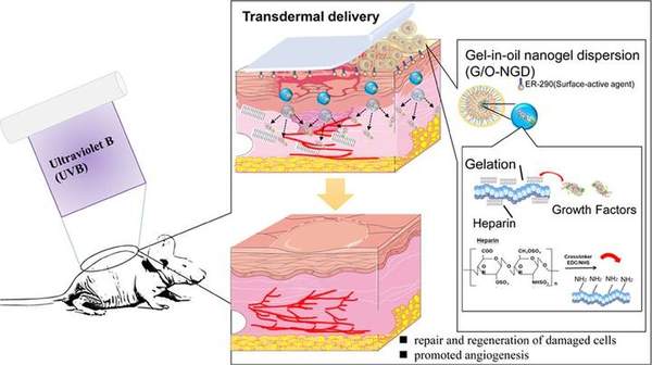
Through cell and mouse experiments, we develop and evaluate protective agents against radiation dermatitis and related skin damage (see Radiation Dermatitis Protective Agents below).
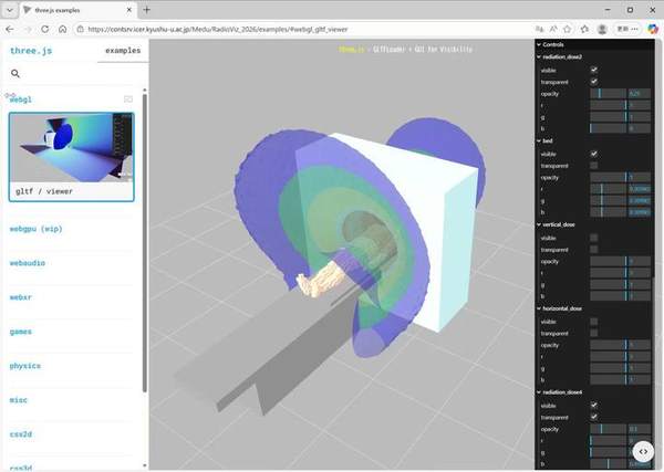
| Monte Carlo simulation | Codes such as PHITS to compute organ doses, scatter distributions and shielding structures — especially for conditions that cannot be measured directly. |
|---|---|
| Dose estimation systems | NCIRF (National Cancer Institute Dosimetry System for Radiography and Fluoroscopy) and related tools for organ and effective dose estimation. |
| Measurement & phantoms | Dosimeters and anthropomorphic phantoms, including in-house phantoms 3D-printed to match CT numbers. |
| Scatter visualization | Our own X-SERVE system for visualizing scatter distributions, plus real-time imaging with a high-sensitivity CMOS camera and pinhole collimator. |
| XR & 3D technology | AR/MR on head-mounted displays such as HoloLens, 3D models viewable in a web browser, and depth cameras for tracking operator position. |
| 3D fabrication | 3D-printed human phantoms and solid models of scatter dose distributions. |
Issues frequently raised in clinical practice, seen from a research perspective
In its 2011 Seoul Statement (formally published as ICRP Publication 118, 2012), the ICRP lowered the equivalent dose limit for the lens of the eye to 20 mSv per year (averaged over five years, not exceeding 50 mSv in any single year). The new limit has applied in Japan since April 2021. For physicians, nurses and radiologic technologists working in IVR and angiography, the head and neck are close to the source and exposure is highly non-uniform, so trunk-level monitoring alone cannot properly estimate the lens dose. We study the actual state of eye lens exposure (see the survey of eye-lens exposure on Basics) and develop site-specific dose evaluation and monitoring methods.
Where protective aprons are worn, non-uniform exposure is assessed by wearing two dosimeters — inside the apron (trunk) and outside at the collar. In practice, however, compliance and operation vary widely between institutions. As a research group of the Japanese Society of Radiological Technology chaired by Fujibuchi, we have surveyed hospitals nationwide on non-uniform exposure management and propose more effective monitoring schemes.
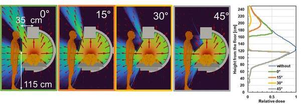
Lead aprons, protective eyewear, ceiling-suspended shields and protective curtains only work when correctly positioned. Combining measurement with Monte Carlo simulation, we quantify the reduction achieved by each device, position and angle, and examine optimal use including where the operator stands.
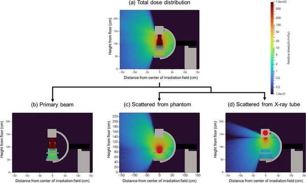
The main source of scatter is the patient's body. Because X-rays scatter in all directions from the point of incidence, the distribution changes markedly with C-arm angle, field size and tube voltage. We visualize the position and direction of scatter sources in three dimensions and implement them as AR and VR learning materials.
In CT, dose optimization builds on size-adjusted evaluation using SSDE (Size-Specific Dose Estimate) and comparison against Diagnostic Reference Levels (DRLs). We combine image-quality assessment (MTF, noise, task transfer function) with dose evaluation to identify settings that reduce exposure while preserving diagnostic quality.
To ensure that statutory training is more than a lecture to sit through, experiential learning with XR materials is effective. We not only develop such materials but also verify their learning effect, pursuing teaching methods that actually change behaviour.
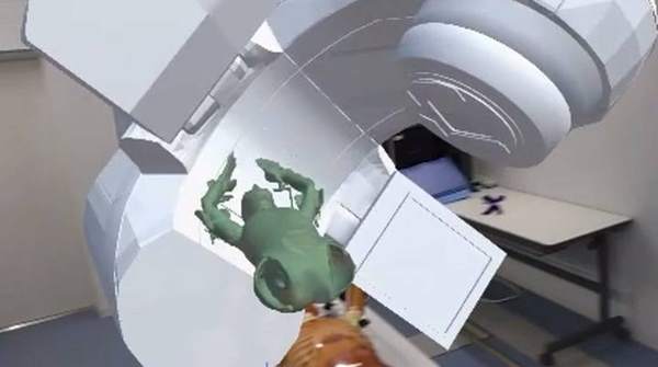
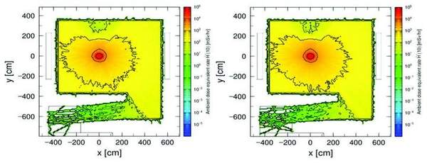
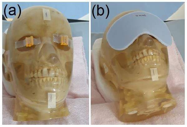
In radiation therapy, we combine XR (AR/VR) with numerical simulation: AR (HoloLens)-based patient setup support, a VR patient experience, and evaluation of photon/neutron ambient dose in the linac room using the Monte Carlo code PHITS and measurements (glass dosimeters, neutron track detectors).
Scattered-radiation distributions obtained by Monte Carlo simulation are implemented as an AR app for iPad/iPhone ("X-SERVE"), tracking staff with LiDAR to estimate dose at multiple points. We visualize how scatter direction changes with C-arm angle and shield placement, and in cardiac angiography we visualize scatter sources in 3D to identify high-dose-rate regions. We are also developing a scatter-visualization camera combining a pinhole collimator, a CMOS camera and a depth camera to image scatter sources in real time.
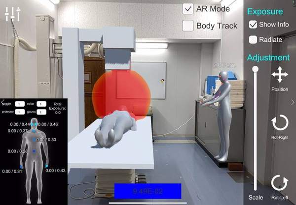
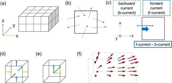
The features, variants and screen examples of the AR app "X-SERVE" are collected on Education — X-SERVE.
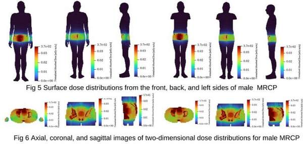
Using mesh-type reference computational phantoms (MRCP) and CT images, we accurately estimate organ and effective doses in CBCT and X-ray examinations. Combined with measurements using real equipment and phantoms, we verify estimation accuracy and aim at patient-specific dose evaluation.
Our laboratory has developed a ground contamination dose simulator that uses the Monte Carlo method to calculate the dose distribution from ground surfaces contaminated with Cs-137, and uses it in lectures and training. The ideas behind environmental protection and decontamination are explained in Basics — ③ public and environmental protection.
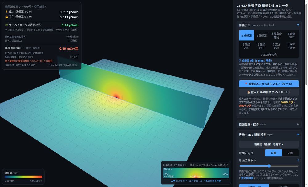
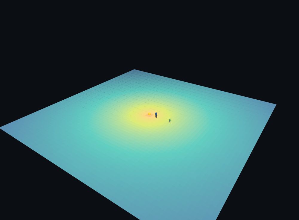
For a point source the inverse-square law applies, so raising the evaluation height from 1 m to 2 m and 4 m lowers the dose to 1/4 and 1/16. Over widely contaminated ground (an area source), the same increase in height lowers it only to 0.85 and 0.69 times.
Calculating which parts of the ground contribute to the dose received by a person standing 1 m above it shows that half comes from within a radius of about 10 m, and 90% from within about 90 m (surface deposition of Cs-137, source radius 400 m).
| Radius band | Area | Contribution | Cumulative |
|---|---|---|---|
| 0–1 m | 3 m² | 7.8% | 7.8% |
| 1–5 m | 75 m² | 28.6% | 36.4% |
| 5–10 m | 236 m² | 14.3% | 50.7% |
| 10–20 m | 942 m² | 13.6% | 64.3% |
| 20–50 m | 6,597 m² | 16.6% | 80.9% |
| over 50 m | 117,810 m² | 19.1% | 100% |
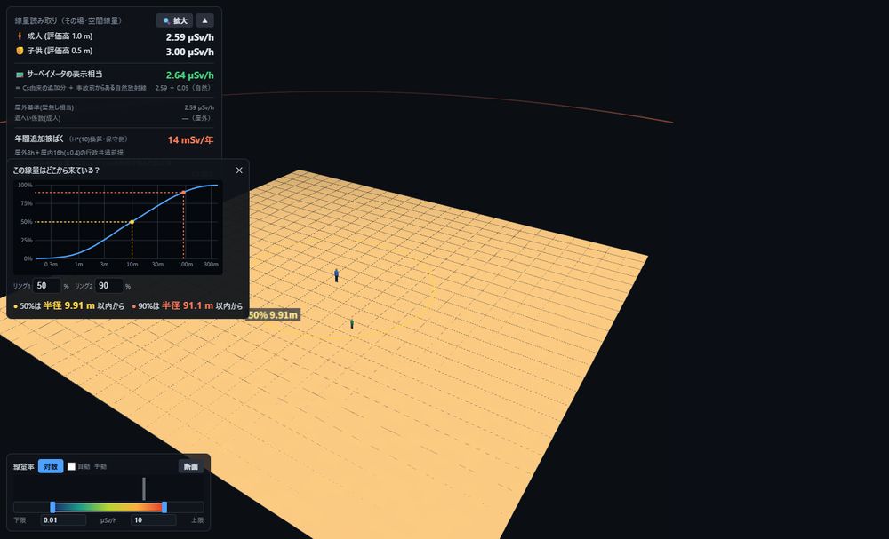
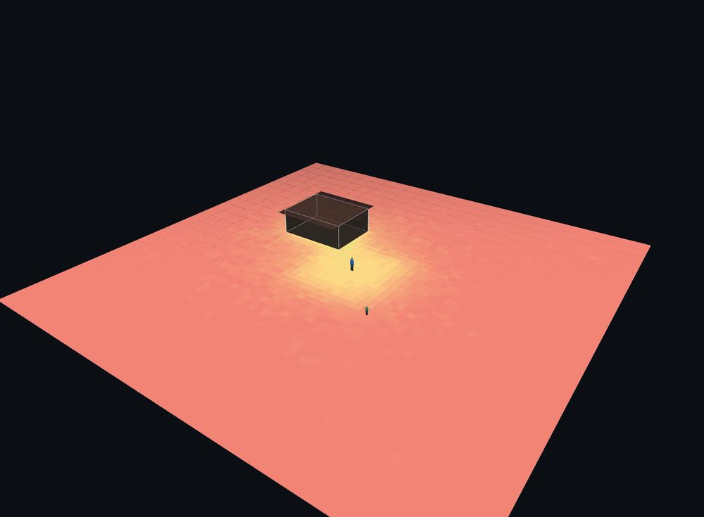
Knowing this distribution makes it possible to estimate quantitatively how effective decontamination will be.
| Decontaminate a 10 m × 10 m garden | about 36% reduction |
|---|---|
| Extend to 20 m × 20 m | about 48% reduction |
| Extend to 50 m × 50 m | about 64% reduction (the ground outside remains contaminated, so the dose does not fall further) |
| Go inside a wooden house | outdoors 0.49 µSv/h → indoors 0.33 µSv/h (about ×0.67) |
All of these calculations use the Monte Carlo method — following the histories of many photons with random numbers and averaging the results. It handles problems involving complex geometry and scattering by statistical sampling rather than analytical formulas, and it is the same tool we use to design shielding for medical facilities and to evaluate scatter distributions (the method is explained in the glossary article "Monte Carlo simulation" (in Japanese)).
Open the simulator in your browser
The values shown are model calculations, not measurements at any specific location.
In radiation therapy, many patients with head-and-neck or breast cancer develop acute radiation dermatitis (ARD) at the irradiated site. Because severe cases can interrupt treatment, we develop new protective agents — such as a film-forming emulsion (FFE) applied to the skin — and evaluate their efficacy through mouse and cell-culture experiments.
The ideas behind our research, the teaching materials that grew out of it, and our publications are presented on the following pages.
The ICRP principles; medical, occupational and public exposure; and radiation protection culture — the ideas our research builds on.
Teaching materials that grew out of our research, how we test them in class, videos, and our browser-based web materials.
About the lab, our members, and how to join as a graduate or international student.
Commissioned shielding and dose evaluation, training for medical staff, and international collaboration.
Please email us directly. We usually reply within a few working days.
For prospective undergraduate, master's, doctoral and international students.
Email about joining the labDose evaluation, shielding calculation, scatter visualisation and XR materials.
Email about collaborationRadiation protection training and FD seminars for medical staff.
Email about training