Our research rests on two pillars: “radiation protection, monitoring, and radioactive waste of patients and staff in medical radiation use” and “visualization of radiation.” Combining experimental measurement, numerical simulation, and field surveys, we propose evidence-based strategies for dose reduction.
1. Assessment of Medical and Occupational Exposure
We quantitatively assess the doses received by patients and healthcare workers in radiological procedures such as fluoroscopy/IVR (image-guided intervention), nuclear medicine, and radiation therapy. We focus on the management of occupational exposure, which has grown in importance since the 2011 recommendations of the ICRP.
2. Dose Evaluation and Monitoring
Using various dosimeters and phantoms, we perform measurements and advance monitoring methods for radiological staff. We characterize site-specific exposure including the lens of the eye and extremities, and propose more effective monitoring schemes.
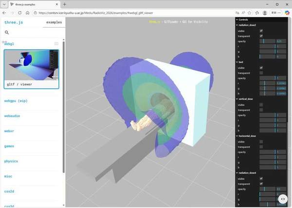
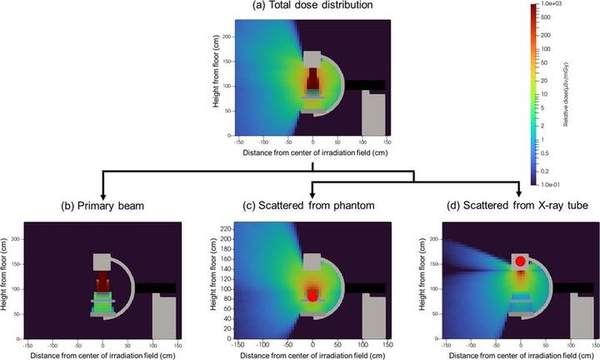
3. Dose-Distribution Analysis by Monte Carlo Simulation
Using Monte Carlo methods, we compute radiation scatter and dose distributions to optimize protective equipment and shielding placement and to quantify dose-reduction effects, validating the simulations against measurements.
4. Safe Management of Radioactive Waste
We study the appropriate management and safety assurance of radioactive waste generated in medical and research settings, contributing to improved safety management in cooperation with the Center for Integrated Radiation Safety Management.
5. Radiation Visualization and XR Material Development
We visualize “invisible” radiation using AR/VR/MR (XR) and implement it as materials for radiation protection education. We develop DX-based dose-reduction programs and verify their educational effectiveness (see the Education / XR page).
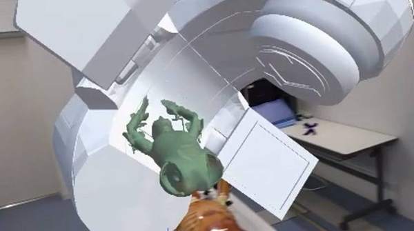
Radiation Therapy Research
In radiation therapy, we combine XR (AR/VR) with numerical simulation: AR (HoloLens)-based patient setup support, a VR patient experience, and evaluation of photon/neutron ambient dose in the linac room using the Monte Carlo code PHITS and measurements (glass dosimeters, neutron track detectors).

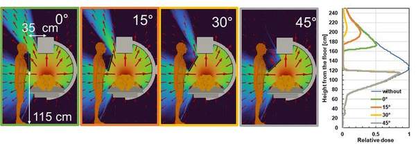
Scatter Visualization & Real-time Imaging
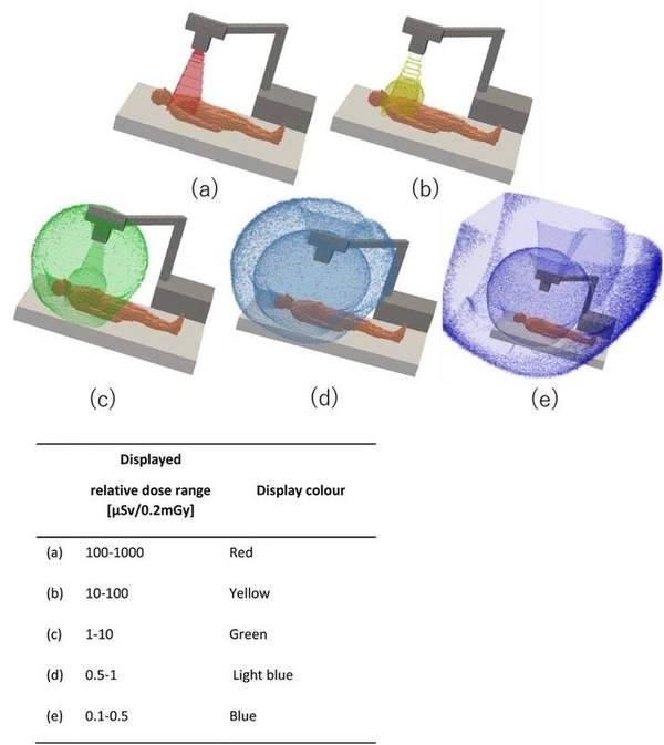
Scattered-radiation distributions obtained by Monte Carlo simulation are implemented as an AR app for iPad/iPhone (“X-SERVE”), tracking staff with LiDAR to estimate dose at multiple points. We visualize how scatter direction changes with C-arm angle and shield placement, and in cardiac angiography we visualize scatter sources in 3D to identify high-dose-rate regions. We are also developing a scatter-visualization camera combining a pinhole collimator, a CMOS camera and a depth camera to image scatter sources in real time.
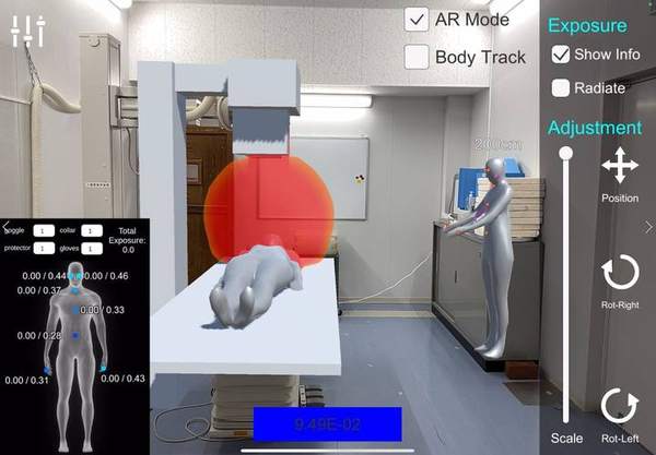
The AR app “X-SERVE” (X-ray examination Scattered Radiation Visualize application) overlays the Monte Carlo scatter distribution on real space on an iPad/iPhone. It shows the 3D spread of scattered radiation in colors by dose level (red, yellow, green, light blue, blue), so staff can intuitively grasp exposure at any position relative to the equipment.
Precise Dose Evaluation for Medical Exposure
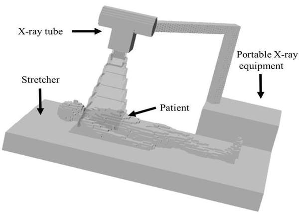
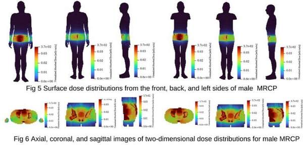
Using mesh-type reference computational phantoms (MRCP) and CT images, we accurately estimate organ and effective doses in CBCT and X-ray examinations. Combined with measurements using real equipment and phantoms, we verify estimation accuracy and aim at patient-specific dose evaluation.
Radiation Dermatitis Protective Agents
In radiation therapy, many patients with head-and-neck or breast cancer develop acute radiation dermatitis (ARD) at the irradiated site. Because severe cases can interrupt treatment, we develop new protective agents — such as a film-forming emulsion (FFE) applied to the skin — and evaluate their efficacy through mouse and cell-culture experiments.
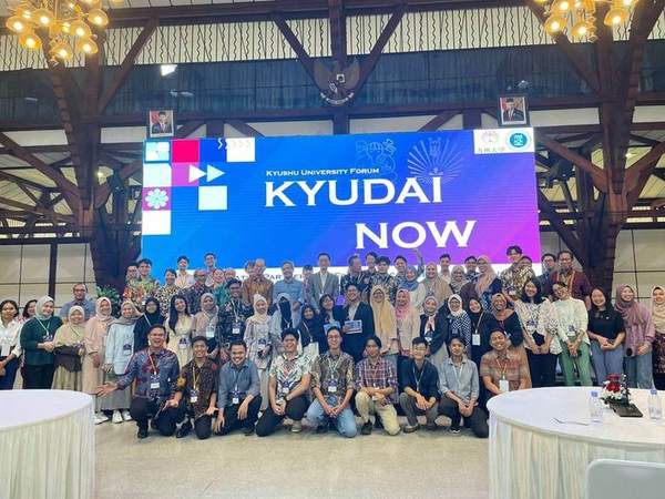
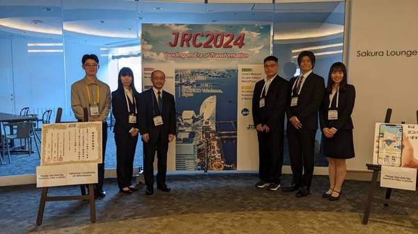
International Collaboration
We collaborate with universities and institutions in Indonesia (Diponegoro University) and Korea (Kangwon National University), advancing radiation protection and medical physics research internationally through joint studies, conferences and mutual visits.
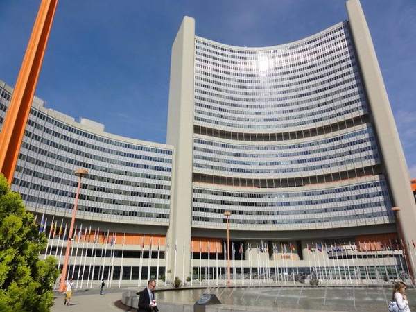
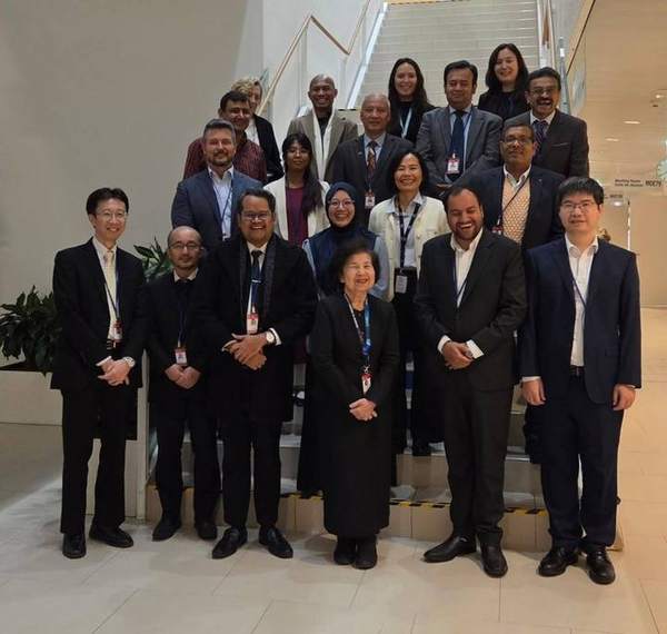
We also participate in an IAEA Regional Cooperative Agreement (RCA) project on strengthening diagnostic/IVR medical physics (2024–2027) together with 16 Asia-Pacific countries, and attended its mid-term review meeting at the IAEA headquarters in Vienna in December 2025.